Introduction
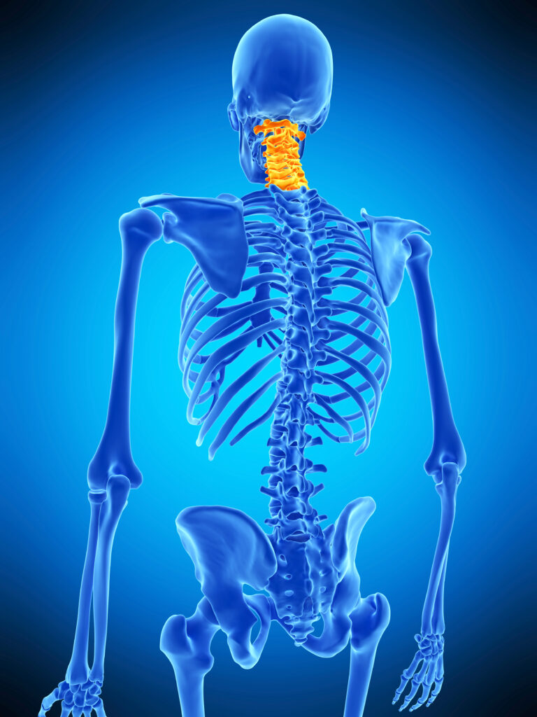
The neck (cervical spine) is made up of 7 bones (C1 vertebra-C7 vertebra) that join the head at the top with the thoracic spine at the bottom. From the 2nd vertebra down there are discs inbetween the bones of the spine which act as shock absorbers and contribute to the flexibility of the neck.
The cervical spine has an important mechanical function in providing strong support for the head whilst also allowing for a good range of head and neck movements.
The cervical spine also has the role to protect the spinal cord that relays information back and forth between the brain and the rest of the body. This is important for muscle power, sensation and other functions such as bladder, bowel and sexual function.
Any problem affecting the cervical spine can cause neck pain, and even neurological problems. The most common problem is degenerative disease, ie wear and tear as part of the normal ageing process. This occurs in the hips, knees, back and can also occur in the neck.
The cervical spine can also be affected by infection, tumour and trauma although these are less common.
The video to the right, produced by the team at Musgrove Park, provides further information about common problems with the Cervical Spine.
Below are sections on specific conditions that can affect the Cervical Spine and also operations of the Cervical Spine
Cervical Spine Conditions
Overview
The whole spine is susceptible to age-related degenerative changes. Your doctor may refer to this as wear and tear.
The vertebrae are separated by intervertebral discs, that act as shock absorbers during loading of the spine that occurs in daily life. Repeated loading over time can lead to the disc becoming dehydrated and degenerate. The disc is then less efficient as a shock absorber and that segment of the spine becomes more susceptible to abnormal loading and further degeneration.
Degenerative changes can lead to disc prolapses (herniation), spurs (bony overgrowth), and loss of disc height which in turn can sometimes cause pressure on the nerves exiting the spinal canal or narrowing of the spinal canal itself.
There may be some activities that reliably trigger flare ups of neck pain and these should be avoided. Maintaining a good posture and staying active are critical for long term spinal health.
Overview
This describes a situation where the wear and tear in the neck causes compression of the spinal cord. This tends to be a chronic condition (when symptoms progress slowly over time), although some patients report a more rapid evolution of symptoms over a course of weeks.
Patients describe a deterioration in hand function, especially their manual dexterity. For example, they may find they are dropping objects, having difficulties with doing buttons, or feel more clumsy with their hands when doing activities that require fine movements. They may also describe a deterioration in their walking, with poor balance. This may be associated with pain and pins and needles in the arms, particularly in the hands.
Cervical myelopathy is an important problem to diagnose as it is caused by spinal cord damage. Any injury to the spinal cord is not necessarily reversible, meaning early diagnosis and treatment is important for preservation of function. The other consideration is that if not treated the symptoms of myelopathy usually progress over time. It is difficult to predict how fast symptoms get worse and this can vary.
The only treatment option for cervical myelopathy is surgery. For patients with moderate or severe symptoms, surgery is recommended. For patients with only mild symptoms there is the option of waiting to see if the situation stabilises.
Overview
This describes a situation when the wear and tear at one or more levels in the neck causes compression of nerve roots as they leave the spinal cord. This may be caused by an acute disc prolapse, or by a more chronic overgrowth of bone.
Patients describe a predominant arm pain, that often radiates from the neck down the arm and sometimes below the elbow into the hand. The pain can be made worse by certain head movements. There is sometimes associated pins and needle and numbness in the arm and hand. Rarely there is an associated weakness in the arm.
In the majority of cases this is a self-limiting condition that improves over 6-12 weeks as the disc prolapse resolves. During this time there is the option of specialised pain relief and sometimes there is a role of an injection of steroids around the compressed nerve in the neck. Sometimes if multiple levels appear affected or when clinical findings don’t correspond with imaging, patients may need further diagnostic tests.
If the arm pain is severe, not controlled with painkillers and persists for more than 3 months there may be a role for surgery.
Cervical Spine Operations
Overview
Surgery from the back of the neck is called a posterior cervical decompression.
Surgery is done under a general anaesthetic. We make an incision at the back of the neck and move muscle away to expose the back of the spine.
We use Xrays to check we are at the correct level. We then remove the bone and soft tissue that is causing spinal cord or nerve root compression with microinstruments under a microscope. At the end of the procedure the spinal cord and nerve roots have been completely decompressed. We then allow the muscles to come back into their natural position and perform a layered closure of the surgical wound.
Occasionally, it is important to fix the spine at the same time. We place screws into the remaining bone and link these to rods to provide an internal brace or scaffold. This provides posterior column support and ultimately promotes bony fusion between the vertebra.
Overview
Surgery from the front of the neck is called an anterior cervical discectomy and fusion (ACDF).
Surgery is done under a general anaesthetic. We make an incision on the side of the neck and develop a plane between the carotid sheath on one side and the trachea and oesophagus on the other. This takes us directly to the front of the spine.
We use Xray’s to check we are at the correct levels. We then remove the entire disc using microinstruments under a microscope. At the end of the procedure the spinal cord and nerve roots have been completely decompressed.
Finally, we place an interbody spacer known as a cage packed with bone graft into the disc and bone space, and we may fix in place with a plate and screws. This provides anterior column support, maintains the height of the space through which the nerve roots are coming out of the spine and ultimately promotes bony fusion between the vertebra.
Videos
This film provides further information regarding an Anterior Cervical Discectomy and Fusion (ACDF).
In this film Anterior Cervical Discectomy and Fusion (ACDF) surgery: post-operative care we talk through the important recovery stages and tips following your ACDF surgery.
Overview
Approaches to the front and the back of the spine have general risks of surgery that include wound complications, infection and bleeding.
With the front (anterior) approach there are risks related to the approach that include hoarseness in the voice and swallowing difficulties. These are very common, but almost always mild and temporary and do not require any treatment. Rarely, they can persist for longer and may require input from a speech and language therapist.
With the back (posterior) approach there is a slightly increased risk of post operative pain in the neck and also an increased risk of infection. Some of the patients who require multiple levels decompressed from the back may sometimes develop over time an abnormal shaped curvature of the neck (kyphosis).
With both approaches there is a risk of cerebrospinal fluid (CSF) leak. This does not normally have any long-term consequences but would need to be repaired at the time of surgery. This can slow your discharge from hospital.
With both approaches there is a risk of an injury to the spinal cord or nerve roots that can lead to arm pain, weakness, paralysis and even loss of life. This is very rare.
There are risks of instrumentation that may require revision surgery, including a small risk of vascular injury that may lead to a stroke.
Finally there is a risk of adjacent level disease (accelerated wear and tear at the level above or below the level of a fused segment) in the longer term, also requiring further surgery.
Videos
In this film pre-operative exercise and balance, our physiotherapist Patrick talks through some key pre-operative exercises. Please complete as prescribed by your physiotherapist.
Further Information (External Links)
Further information on Cervical Spine Conditions:-
Further information on Cervical Spine Surgery:-